Achondroplasia In Children: Everything You Need To Know
Achondroplasia is one of the most common forms of dwarfism that we can observe in the world population. For every 25,000 babies born, one of them suffers from this genetic condition that affects the bone system.
According to some research, 75% of achondroplasia cases in children are due to sudden mutations in genes, which take place during fetal development. On the contrary, in the remaining 25% of cases it is a hereditary problem, transmitted by the genetic background of the parents. This genetic mutation causes approximately 70% of the total cases of dwarfism.
Achondroplasia in children is classified as a disproportionate type of dwarfism (DSS). This condition affects the normal development of the long bones, while there is a development of the normal length of the spine. That is why disproportionate growth of body structure occurs in affected individuals.
In children, achondroplasia triggers an alteration in cellular DNA, affecting a certain growth factor (receptor 3), present in fibroblasts, which are the typical cells of connective tissue. This leads to an abnormality in cartilage formation and a subsequent block in normal bone growth. It poses a high risk of death in the infant stage as spinal compression or obstruction of the respiratory system may occur.
Probability of achondroplasia in children
The odds of a child with healthy parents suffering from this disease is one in 25,000. While it increases by 50% if one parent has the condition, it increased to 75% if achondroplasia is present in both. In addition, there is a 2.5% chance of having homozygous achondroplasia if both parents are affected.
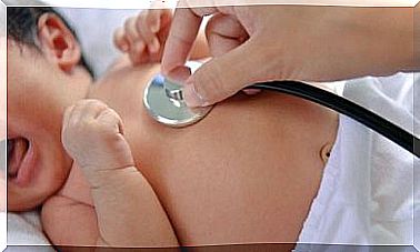
How to diagnose achondroplasia in children
Symptoms can be assessed with an antenatal diagnosis, to be made during the fetal development phase. The presence or absence of the disease can be deduced from bone measurements, but it is only after birth that it can be diagnosed in complete safety.
Treatment for achondroplasia in children
This is a condition for which there is no drug treatment that can reverse the mutation. Despite this, it was possible to locate the gene that acts as a transmitter on chromosome 4.P and 16.3, known as the “short arm of chromosome 4”.
Intellectual capacity and physical characteristics
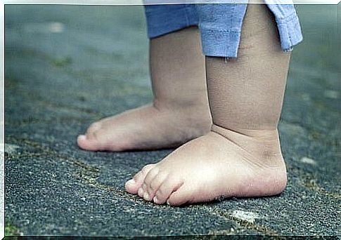
Children with achondroplasia develop normal intellectual ability. On the other hand, the main problems concern the final size of the child affected by the disease. In fact, once it reaches adulthood, it can reach a maximum height of just 120 cm.
Long bones such as the femur and humerus are shorter, comparing the forearm (radius and ulna) and the lower leg. The size of the head is larger than the size of the body, with a large forehead and a particularly flattened nasal septum. The legs can be arched, a condition that can cause a lot of pain, greatly affecting the individual’s motor skills.
In some cases, problems with the teeth may be recorded, which can grow incorrectly. In general, the feet are wider, with narrow toes and the sole of the foot flat. Muscle tone is weak and children suffer from slower development right from the start. They can count on what is known as the “trident hand”, which shows a larger space between the middle and ring fingers.

Other conditions associated with achondroplasia
In addition to physical characteristics, children with achondroplasia may have some diseases associated with the suboptimal development of their body. Fortunately, however, with due attention, they will be able to lead a normal life into adulthood.
- Apnea : the individual stops breathing for a few seconds.
- Otitis : Children with achondroplasia often suffer from ear infections, and if left untreated, they can cause serious damage.
- Spinal cord compression : This occurs when the canal that connects the head with the spine is very small, thus compressing the cord causing breathing problems.
- Hydrocephalus : some fluid accumulates in the baby’s brain, it can be noticed by an abnormal and rapid increase in the size of the head. Therefore, head measurements will need to be monitored regularly.
- Kyphosis : This is a small curvature that forms on the back, at the top. It usually occurs before the baby starts walking and then disappears.
- Lordosis : Another curvature that occurs in the lower back, inward. It usually occurs after the baby starts walking and can be treated with postural gymnastics.